
a luxury Mammamogram experience for the Springfield, mo area
3-D Mammogram
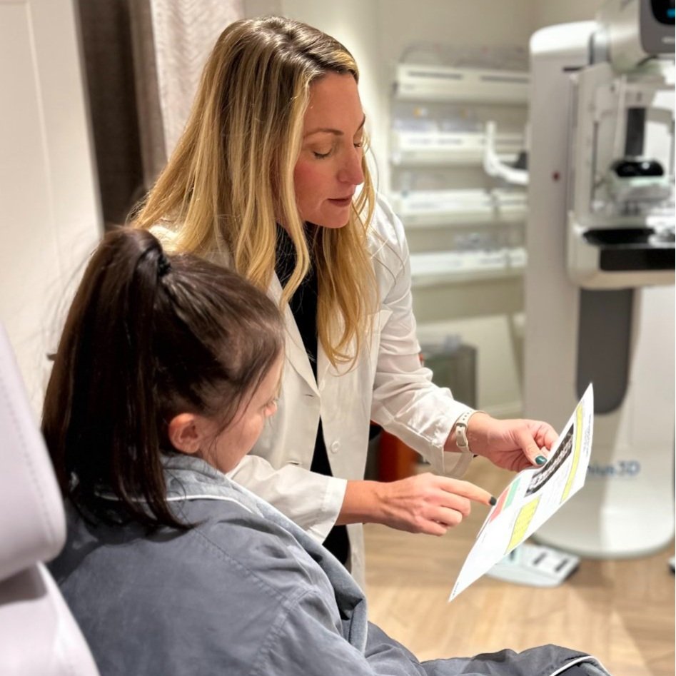
A 3D mammogram (breast tomosynthesis) uses multiple breast X-rays to create a three-dimensional picture of the breast. The technologically advanced 3D Mammogram by Hologic called “Genius 3D” is utilized for all screening and diagnostic mammograms at BIO. A screening mammogram is used to look for breast cancer in women who have no new breast issues. A diagnostic mammogram is performed to evaluate areas of concern on a screening mammogram, or for those presenting with new breast issues that may signal underlying breast cancer such as a lump, nipple discharge, or certain skin changes.
Tomosynthesis-guided biopsy
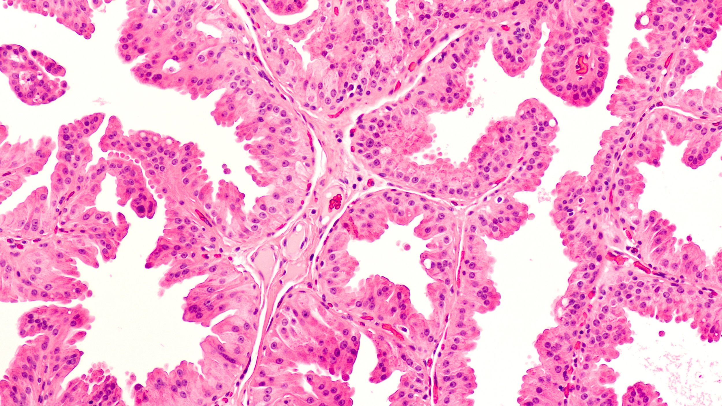
A breast biopsy is performed to remove tissue from a suspicious area in the breast, which is then examined by a pathologist under a microscope to determine a diagnosis. Tomosynthesis-guided Breast Biopsy is not designed to remove the entire lesion. However, the goal is to remove enough tissue to allow the pathologist to make a diagnosis.
In a tomosynthesis-guided breast biopsy, the radiologist uses 3-D mammographic imaging to guide the biopsy device to the site of the abnormality. Tissue samples are obtained, and a tiny marker is placed at the biopsy site to mark where the tissue was sampled. This will be visible on future mammograms and if surgery is needed, the marker will serve as a guide to direct the surgeon to the area that needs to be removed.
Breast Ultrasound
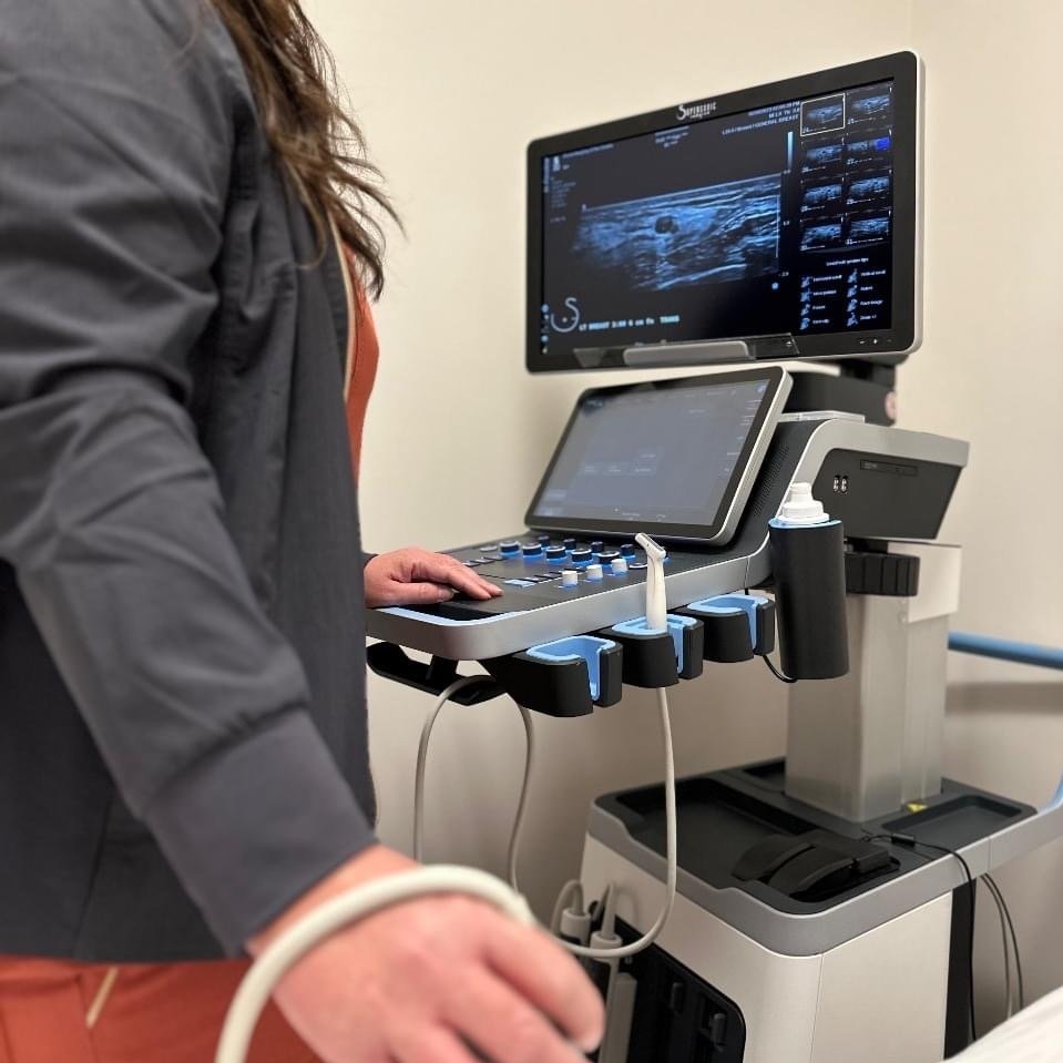
A breast ultrasound uses high-frequency sound waves to a create a picture of the tissues inside the breast. The sound waves pass through the breast and bounce back, or echo, from various tissues to form a picture of the internal structures. It is not invasive and involves no radiation or X-rays. The breast ultrasound can show all areas of the breast, including the area closest to the chest wall, which can be hard to study with a mammogram.
At BIO, we perform both diagnostic and whole breast screening ultrasounds.
Bone Densitometry
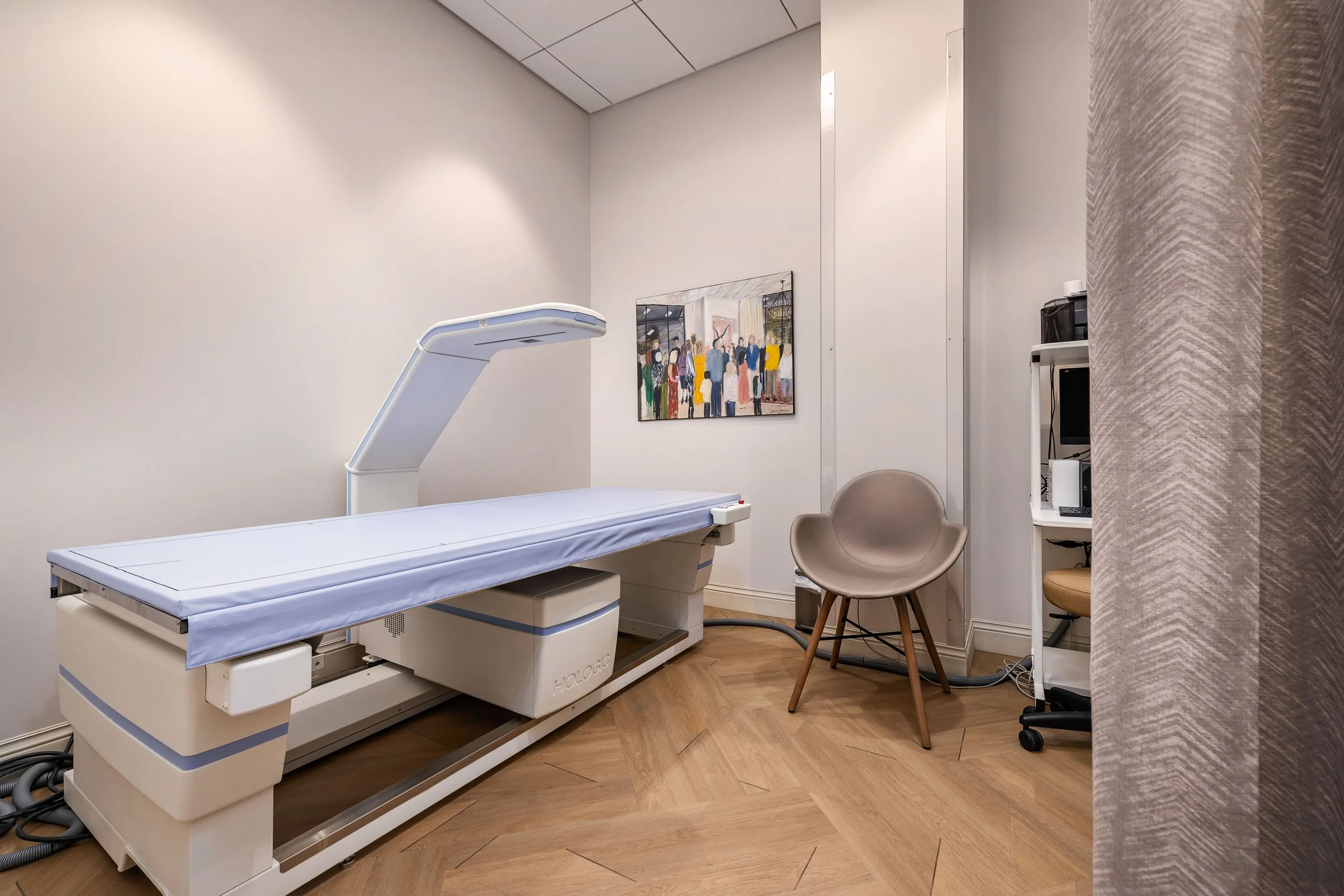
Bone density scanning, also called dual-energy x-ray absorptiometry (DEXA) or bone densitometry, is an enhanced form of x-ray technology that is used to measure bone loss. DEXA is today’s established standard for measuring bone mineral density (BMD).
DEXA bone density scan provides an estimate of the strength of your bones. DEXA can measure as little as 2% of bone loss per year.
DEXA is used to diagnose osteoporosis, a condition that often affects women after menopause, and can assess a person’s risk for developing fractures. The exam is fast, comfortable, and uses very low doses of radiation.
Body composition
Dual X-ray Absorptiometry (DEXA) is one way to determine our body composition. DEXA has become the gold standard in body composition measurement and tracks changes in muscle mass and body fat using X-ray technology. A DEXA scan for body composition generates a personalized report outlining how much fat, muscle mass and bone you have in your body, allowing you to measure progress when implementing changes such as adopting a weight loss program or taking up resistance exercise to grow your muscle mass.

Ultrasound guided biopsy and aspiration
A breast biopsy is performed to remove tissue from a suspicious area in the breast, which is then examined by a pathologist under a microscope to determine a diagnosis. An ultrasound-guided biopsy is not designed to remove the entire lesion. However, the goal is to remove enough tissue to allow the pathologist to make a diagnosis.
In an ultrasound-guided breast biopsy, the radiologist uses ultrasound imaging to guide the biopsy device to the site of the abnormality. Tissue samples are obtained, and a tiny marker is placed at the biopsy site to mark where the tissue was sampled. This will be visible on future mammograms and if surgery is needed, the marker will serve as a guide to direct the surgeon to the area that needs to be removed.
Risk Assessment Consultation
At BIO we take an innovative approach to breast cancer screening and prevention that personalizes care to the unique needs of each woman. While annual mammographic screening should begin at age 40 for women of average risk, higher-risk women should start screening earlier and may benefit from supplemental screening modalities. At your Risk Assessment Consultation your personal and family history will be reviewed with a professional to identify genetic and non-genetic factors that may increase your risk of developing breast cancer. The most accurate and up to date risk assessment model will be applied to your personal data and will determine your individual average lifetime risk of developing breast cancer.


Second Opinion
At BIO, we have the expertise and specialized knowledge to provide you and your doctor added confidence in your breast diagnosis. A second opinion is simply having another specialized breast radiology physician review your prior breast imaging, imaging reports, and test results and provide their opinion on your diagnosis. A second opinion may confirm your original diagnosis, or may provide another perspective on it, possibly suggesting additional imaging or procedures to arrive to the correct diagnosis. Even if you’ve already had treatment, it’s not too late to get a second opinion.